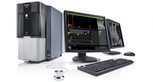
Unlike stereomicroscopes and compound microscopes which use light reflected off or transmitted through a specimen to convey visual information to the observer's eye magnified by a series of lenses, a scanning electron microscope uses an entirely different technique. Specimens are specially prepared by adhering to a stub, dehydrating, and coating with an ultra-thin layer of a conductive metal (usually gold). After being loaded into the SEM, they sit beneath the electron beam in a lightless chamber under vacuum pressure. Electrons continually bombard the specimen with some being absorbed and other bouncing off to be collected by sensors which build a fine-grain image of the specimen's topology on the screen. Microscopic physical features (ultrastructure) are viewable with this technique, but the images are monochrome because no light is used and color is a property light.
Whereas most SEMs are enormous pieces of equipment with complex functionalities that limit use to those with significant training, funding, and research goal, the Phenom SEM is a desktop version that is comparatively user-friendly and accessible for art and design students at RISD to explore a level of the physical world we rarely get a chance to personally tread.
The Phenom allows for magnifications of up to 45,000X (well beyond the practical capability of any type of light microscope) and our collection of prepared specimens, "Nearly Nanoville", affords a diverse array microscopic landscapes to traverse. No two trips to Nearly Nanoville are quite the same!


